F1 doctors must demonstrate competence in 15 procedures to become eligible for full GMC registration. Here are some helpful tips on performing intramuscular injections, performing and interpreting ECGs, and performing and interpreting peak flow.
Performing intramuscular injections
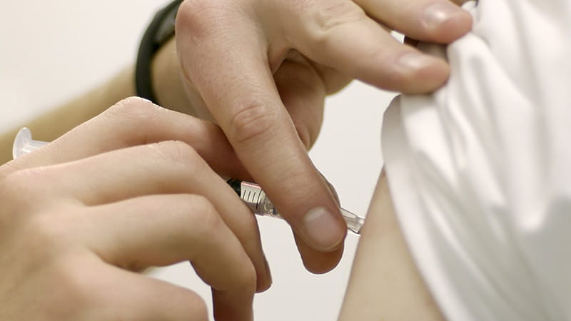
I first performed this skill on my complex care placement, administering naloxone to a patient with an opiate overdose. IM injections are typically given by nurses (politely ask to be shown how), so once I prescribed it, I asked the nurse to allow me to administer it.
I set up the equipment using good aseptic non-touch technique (ANTT), starting with washing my hands and drawing up the medication.
There are two common sites for IM injections to be administered. The middle-third of the deltoid is generally used for small injections in patients who are not cachectic. Alternatively, the upper outer quadrant of the gluteal region may be used. To visualise this, I imagined the midpoint of a line drawn from the greater trochanter of the femur to the posterior superior iliac spine. The injection site should be slightly superior and lateral to this point to minimise the risk of damaging the sciatic nerve.
I stretched the skin taut and inserted the needle at a 90 degree angle, holding it like a pen and warning the patient of a sharp scratch. I drew back before injecting to ensure that I hadn’t hit a vessel by checking no blood was aspirated. I then injected slowly and made sure the patient was comfortable. Once I completed the procedure, I signed and documented it in the drug card.
Performing and interpreting ECGs
My first attempt at performing an ECG came during an on-call shift on a patient with tachycardia who turned out to be in fast AF. ECGs can be difficult to interpret so it is useful to discuss them with a senior if you are unsure. As with any investigation, I ensure I compare it against previous ECG traces for that patient – this lets me know if the LBBB or T-wave inversion I’m seeing is new or not.
When performing ECGs the patient’s chest must be exposed so I would always draw the curtains to maintain the patient’s privacy and dignity. The ECG leads are comprised of six chest leads and four limb leads. Correct lead positioning is extremely important in producing a good quality ECG. In females, I have learned it is preferable to place the electrodes under the breasts rather than overlying them.
Before carrying out the procedure, I entered the patient details into the ECG machine, and inspected the screen to see whether all traces are clearly visible. I have found common artefacts which may be encountered include patient movement leading to a ‘wandering baseline’ and chest lead reversal causing abnormal progression in the R-wave size from V1 to V6.
I was taught a simple systematic approach to interpret ECGs: rate; rhythm and the presence of P-waves before every QRS complex; axis; QRS morphology including BBB; ST elevation/depression; and T-wave morphology. Use a systematic approach with which you are familiar. A structured approach can help your ECG interpretation and facilitates good documentation. I have learned that ECG traces should be interpreted in the context of a given patient’s clinical status to minimise the risk of missing anything pertinent.
Performing and interpreting peak flow
Peak flow assessments are a simple non-invasive test that can be performed quickly at the bedside. You can get this signed off early in your FY1 year. They are most commonly performed on respiratory wards and in pre-op clinics.
I have found that abnormal readings are often due to poor technique rather than pathology but explaining correct technique in a clear and concise manner can help to avoid this. I found that it was best to explain the procedure to the patient and then perform a demonstration with the peak flow meter. The demonstration can also help prevent patients feeling embarrassed when performing the test.
I find it is useful to repeat the test three times and select the best peak expiratory flow rate (PEFR) reading for interpretation. I would then ideally compare these results to the patient’s own PEFR values from within the last two years, but these may be based on predicted values estimated using the patient’s sex and age. You can often access these charts online. I’ve learned that a value less than 50 per cent of previous or predicted values indicates a severe airway obstruction. As before, I would always interpret PEFR alongside my other investigation findings and clinical assessment of the patient.
Completing the 15 procedures is a vital part of completing the FY1 year, so try to be organised and get them signed off as early as possible. Before starting FY1, have a think about which placements would give you the best opportunities to practise each skill. For example, you can often administer local anaesthetic on your surgical placements but rarely in psychiatry.
FY1 is a huge learning curve but with a bit of forward planning and proactivity you can become more confident in your clinical practice and use of skills.
Dr Priya Mistry is a foundation year one doctor based in Bolton
This page was correct at the time of publication. Any guidance is intended as general guidance for members only. If you are a member and need specific advice relating to your own circumstances, please contact one of our advisers.
Read more from this issue of FYi

Save this article
Save this article to a list of favourite articles which members can access in their account.
Save to library